| Headneck Nodal Levels |
| Level 1a |
| Level 1b |
| Level II |
| Level III |
| Level IV |
| Level V |
| Level VI |
| Retropharyngeal Nodes |
| All Pages |
Page 1 of 9
INTRODUCTION
Applied Anatomy
Newer techniques of radiation therapy, specifically Intensity Modulated Radiotherapy (IMRT ), require detailed anatomical knowledge. Drawing the gross tumor volume requires interpretation of Computed tomography scans, Magnetic Resonance Imaging and FDG Positron Emission Tomography scans. Both clinical findings and imaging findings are taken together to define the gross tumor volume. For demonstration click here.
The major challenge for a radiation oncologist is to draw the clinical target volume. The questions that need to be asked prior to drawing CTV are
- What is the pattern of lymphatic spread?
- Which are the nodal levels most commonly involved?
- What is the frequency of involement in each nodal level?
- What are the anatomical boundaries of each nodal level?
- How are these boundaries drawn on computed tomography scans?
The nomenclature used is as decscribed by Robbins, i.e., Levels I to VI. In addition, retropharyngeal nodes,which are not included in Robbins classification, are also described. The anatomical boundaries of nodal levels defined by Robbins are based on surgical procedures and the structures used to define the specific boundaries are not easily recognized on CT scans. The RTOG has published guidelines and an atlas which detail the procedure of drawing nodal levels on CT scans. Detailed anatomical description as per the nodal levels is given below.
LEVEL Ia
Level I is a triangular region which contains the submental nodes. Its boundaries are as under.
Anterior: Platysma muscle and symphysis menti.
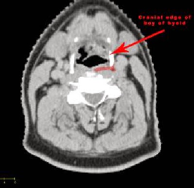
Posterior: Body of hyoid.
Cranial: Geniohyoid muscle or a plane tangent to the basilar edge of the mandible.
Caudal: Hyoid bone.
Lateral: medial edge of the anterior belly of the digastric muscle.
Medial: The medial limit of level Ia is virtual, as the region continues into the contralateral level Ia.
Drainage area for Level Ia
Skin of the chin, the mid-lower lip, the tip of the tongue, and the anterior floor of the mouth.
Level Ia is at greatest risk of harboring metastases from cancers arising from the floor of the mouth, the anterior oral tongue, the anterior mandibular alveolar ridge, and the lower lip.
LEVEL IbIt includes the submandibular nodes. Its boundaries are given below.
Anterior: Platysma muscle and symphysis menti.
Posterior: posterior edge of the submandibular gland.
Cranial: limited by the mylohyoid muscle and the cranial edge of the submandibular gland,
Caudal: Plane crossing the central part of the hyoid bone.
Lateral: basilar edge and inner side of the mandible, the platysma and the skin.
Medial: lateral edge of the anterior belly of the digastric muscle.
Drainage area for Level Ib
Submental lymph nodes, the medial canthus, the lower nasal cavity, the hard and soft palate, the maxillary and mandibular alveolar ridges, the cheek, the upper and lower lips, and most of the anterior tongue.
Nodes in level Ib are at risk of developing metastases from cancers of the oral cavity, anterior nasal cavity, soft tissue structures of the mid-face and the submandibular gland.
LEVEL IILevel II includes the upper jugular lymphnodes situated along the upper third of Internal Jugular Vein and upper portion of spinal accessory nerve. Its boundaries are given below.
Anterior: Anteriorly by the posterior edge of the submandibular gland, the anterior edge of the carotid artery and the posterior belly of the digastric muscle.
Posterior: Posterior edge of the sternocleidomastoid (SCM) muscle.
Cranial: Caudal edge of the lateral process of the first vertebra, which correspond to the insertion of the posterior belly of the digastric muscle to the mastoid which is the surgical landmark.
Caudal: Body of the hyoid bone.
Lateral: Medial edge of the SCM and the platysma
Medial: medial edge of the carotid artery and the paraspinal muscles (levator scapulae and splenius capitis)
Level II is further divided into Level IIa and IIb. the posterior edge of the IJV is taken as the boundary between levels IIa and IIb.
Drainage area for Level II
Level II receives efferent lymphatics from the face, the parotid gland, and the submandibular, submental and retropharyngeal nodes. Level II also directly receives the collecting lymphatics from the nasal cavity, the pharynx, the larynx, the external auditory canal, the middle ear, and the sublingual and submandibular glands.
The nodes in level II are therefore at greatest risk of harboring metastases from cancers of the nasal cavity, oral cavity, nasopharynx, oropharynx, hypopharynx, larynx, and the major salivary glands. Level IIb is more likely associated with primary tumors of the oropharynx or nasopharynx, and less frequently with tumors of the oral cavity, larynx or hypopharynx.
LEVEL III
Level III contains the middle jugular lymph nodes located around the middle third of the IJV. It is the caudal extension of level II. Its boundaries are given below.
Anterior: Posterolateral edge of the sternohyoid muscle and the anterior edge of the SCM muscle.
Posterior: Posterior edge of the SCM muscle.
Cranial: Caudal edge of the body of the hyoid bone.
Caudal: Caudally by the caudal edge of the cricoid cartilage.
Lateral: Medial edge of the SCM muscle.
Medial: Medial edge of the internal carotid artery and the paraspinal muscles (scalenius).
Drainage area for Level III
Level III contains a highly variable number of lymph nodes and receives efferent lymphatics from levels II and V, and some efferent lymphatics from the retropharyngeal, pretracheal and recurrent laryngeal nodes. It collects the lymphatics from the base of the tongue, tonsils, larynx, hypopharynx and thyroid gland [18].
Nodes in level III are at greatest risk of harboring metastases from cancers of the oral cavity, nasopharynx, oropharynx, hypopharynx and larynx.
LEVEL IV
Level IV includes the lower jugular lymph nodes located around the inferior third of the IJV. According to Robbins, it extends from the caudal limit of level III to the clavicle. Its boundaries are given below.
Anterior: Posterolateral edge of the sternohyoid muscle and the anterior edge of the SCM muscle.
Posterior: Posterior edge of the SCM muscle.
Cranial: Caudal edge of the cricoid cartilage.
Caudal: Two cm cranially to the cranial edge of the sternoclavicular joint.
Lateral: Medial edge of the SCM muscle.
Medial: Medial edge of the internal carotid artery and the paraspinal muscles (scalenius).
Drainage area for Level IV
Level IV contains a variable number of nodes and receives efferent lymphatics primarily from levels III and V, some efferent lymphatics from the retropharyngeal, pretracheal and recurrent laryngeal nodes, and collecting lymphatics from the hypopharynx, larynx and thyroid gland.
Level IV nodes are at high risk of harboring metastases from cancers of the hypopharynx, larynx and cervical esophagus.
LEVEL V
Level V includes the lymph nodes of the posterior triangle group. Its boundaries are given below.
Anterior: Posterior edge of the SCM muscle.
Posterior: Practically, a virtual line joining the antero-lateral border of both trapezius muscles can be use to set the posterior limit of level V.
Cranial: Cranial edge of the body of the hyoid bone.
Caudal: CT slices encompassing the cervical transverse vessels.
Lateral: Platysma muscle and the skin.
Medial: Splenius capitis, levator scapulae and scaleni (posterior, medial and anterior) muscles.
Drainage area for Level V
Level V receives efferent lymphatics from the occipital and post-auricular nodes as well as those from the occipital and parietal scalp, the skin of the lateral and posterior neck and shoulder, the nasopharynx and the oropharynx (tonsils and base of the tongue).
Level V lymph nodes are at high risk or harboring metastases from cancers of the nasopharynx, oropharynx, subglottic larynx, the apex of the piriform sinus, the cervical esophagus and the thyroid gland.
LEVEL VI
Level VI, also called the anterior neck compartment, contains the lymph nodes located in the visceral space: the pre- and paratracheal nodes including the precricoid (Delphian) node and the perithyroid nodes including the lymph nodes along the recurrent laryngeal nerves. Its boundaries are given below.
Anterior: Platysma and the skin.
Posterior: Separation between the trachea and the esophagus.
Cranial: Caudal edge of the body of the thyroid cartilage.
Caudal: Cranial edge of the sternal manubrium.
Lateral: Medial edge of the thyroid gland, the skin and the antero-medial edge of the SCM muscle.
Medial: Midline structure so no medial boundary.
For the paratracheal and recurrent nodes, the cranial limit is the caudal edge of the cricoid cartilage.
For the pretracheal nodes, the posterior limit is the trachea and the anterior edge of the cricoid cartilage.
Drainage area for Level VI
Level VI receives efferent lymphatics from the thyroid gland, the glottic and subglottic larynx, the hypopharynx and the cervical esophagus.
These nodes are at high risk or harboring metastases from cancers of the thyroid gland, the glottic and subglottic larynx, the apex of the piriform sinus and the cervical esophagus.
RETROPHARYNGEAL NODES
Retropharyngeal lymph nodes lie within the retropharyngeal space, which extends cranially from the base of the skull to the cranial edge of the body of the hyoid bone caudally. Its boundaries are given below.
Anterior: Fascia under the pharyngeal mucosa.
Posterior: Prevertebral m.(longus colli, longus capitis).
Cranial: Base of skull.
Caudal: Cranial edge of the body of hyoid bone.
Lateral: Medial edge of the internal carotid artery.
Medial: Midline.
Drainage area for Retropharyngeal Nodes.
Retropharyngeal node involvement occurs in primary tumors arising from (or invading) the mucosa of the occipital and cervical somites, e.g. of the nasopharynx, the pharyngeal wall and the soft palate. Retropharyngeal nodes are also at risk in case of pharyngeal tumors with positive neck nodes in other levels in the neck.


No comments:
Post a Comment